What Actually Happens at an Echocardiogram
Short answer
An echocardiogram uses sound waves, not radiation, to produce a moving picture of your heart's size, valves, walls and pumping strength. Most people get a transthoracic version (TTE) from outside the chest, 30 to 60 minutes, no preparation needed. A transesophageal version (TEE) threads a small transducer down the throat for a clearer view when needed, and does carry some real risk.
A doctor hears something during an exam, or you've had chest pain, shortness of breath, or swelling that needs an explanation, and the word "echocardiogram" comes up. For most men, that's the extent of the briefing — what actually happens in the room is left unexplained until you're already lying on the table.
This article covers what an echocardiogram actually checks, why your doctor would order one, the real difference between the two main types, how to prepare, what the test physically feels like, and what the risks and results genuinely involve.
What an echocardiogram actually is
MedlinePlus defines an echocardiogram as a test that uses sound waves to create pictures of the heart — more detailed than a standard X-ray image, and without exposing you to radiation. It uses sound waves to check your heart's size and structure, your heart's valves, the strength of your heart's walls, how well your heart pumps blood, and the pericardium, the sac that surrounds the heart.
Unlike a single still image, an echocardiogram shows the heart while it is actually beating — valves opening and closing, chambers filling and emptying, blood moving through in real time. A Doppler echocardiogram specifically evaluates the motion of blood through the heart, useful for spotting leaking or blocked valves.
It can also show up on paperwork under a different name, which is worth knowing before you assume a new test has been ordered. MedlinePlus lists the alternate names: transthoracic echocardiogram (TTE), transesophageal echocardiogram (TEE), stress echocardiography, point-of-care ultrasound (POCUS), contrast echocardiography, strain imaging, 3D echocardiography, echo, heart echo, heart or cardiac ultrasound, and heart sonogram — all referring to some version of the same underlying test.
Why your doctor orders one
MedlinePlus lists specific reasons a provider orders an echo: a heart murmur, damaged heart valves, shortness of breath, edema (swelling in the legs), unexplained chest pain, rheumatic fever, any congenital heart defect, or after a heart attack, stroke, or heart surgery. It is also used to monitor chronic or congenital heart conditions over time, and before and after surgery or treatment for a heart condition.
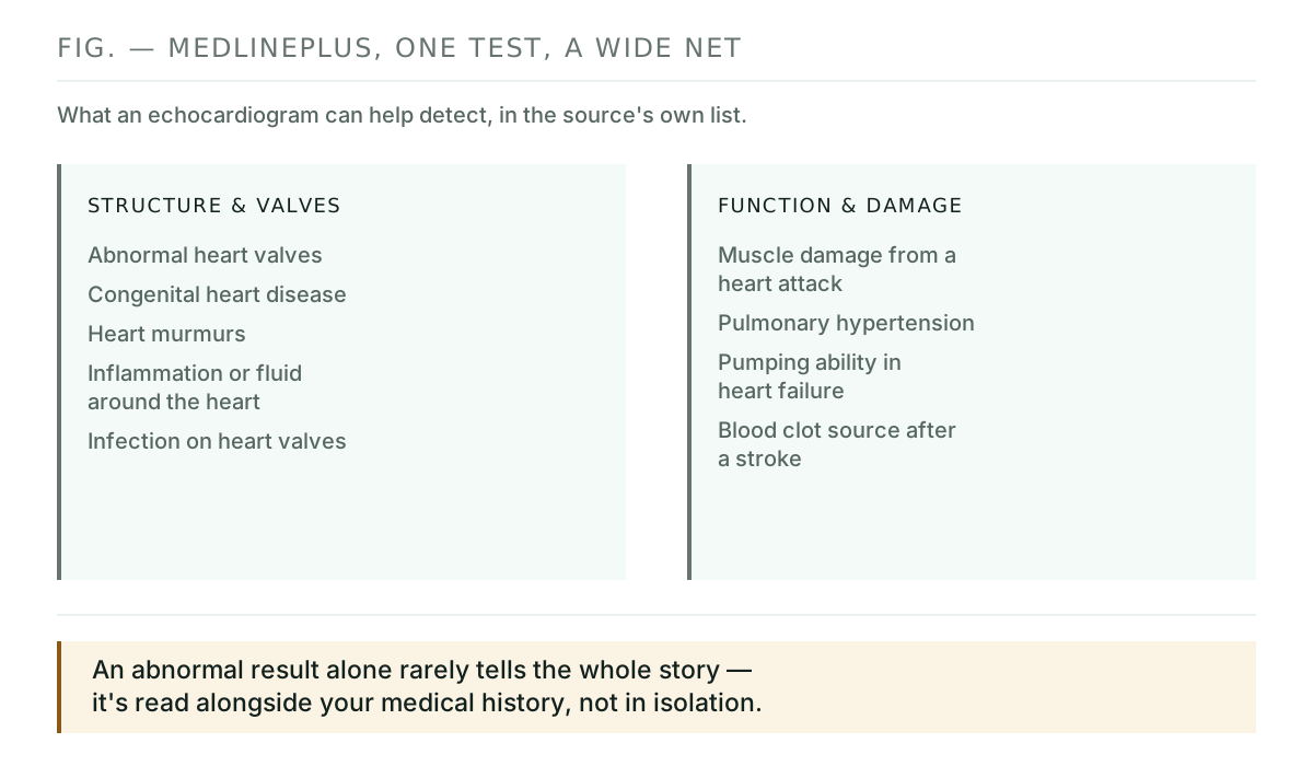
The test can help detect abnormal heart valves, congenital heart disease, damage to the heart muscle from a heart attack, murmurs, inflammation or fluid around the heart, infection on or around the valves, pulmonary hypertension, a blood clot source after a stroke, and simply how well the heart is pumping in people with heart failure.
MedlinePlus also notes that the heart conditions an echo is used to investigate can look different depending on your sex — women are more likely to experience nausea, extreme fatigue, and pain in the neck or back during a heart attack, and certain health conditions raise heart disease risk in women more than men. It's a reminder that the same test gets read against a somewhat different baseline depending on who's on the table.
The two main types: TTE and TEE
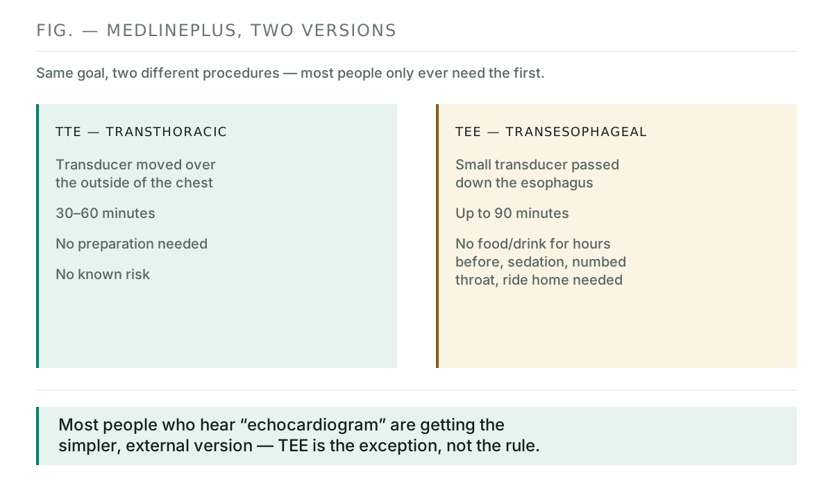
MedlinePlus states that TTE — transthoracic echocardiogram — is the type most people will have. A sonographer places a transducer on various locations on your chest and upper abdomen, and the device releases high-frequency sound waves and picks up the echoes. The echocardiography machine converts those echoes into moving pictures.
A TEE — transesophageal echocardiogram — is different: a long, flexible tube with a small ultrasound transducer at the end is passed down the throat and esophagus, after the back of the throat is numbed. MedlinePlus says this gets clearer images and is used to look for things like infection (endocarditis) or blood clots. Your provider may recommend a TEE specifically if a TTE's images were unclear — which can happen because of chest shape, lung disease, or excess body fat — or if a part of the heart needs a more detailed look.
How to prepare
For a TTE, MedlinePlus says no special steps are needed beforehand. For a TEE, you will not be able to eat or drink for several hours before the test, and because you'll receive a sedating medicine, you'll need someone to take you home afterward — the sedative can leave you drowsy for several hours.
What actually happens during the test
MedlinePlus is specific about who does what: a specially trained technician called a sonographer performs the test itself, and a heart doctor — a cardiologist — interprets the results afterward. The person running the transducer over your chest is usually not the one who tells you what it means, which is worth knowing so you don't expect answers in the room.
For a TTE: MedlinePlus describes removing clothing from the waist up (a gown is provided), lying on an exam table on your back, electrodes placed on your chest to monitor your heartbeat, and a small amount of gel spread on your chest where the transducer is moved. You'll feel a slight pressure from the transducer, and may be asked to breathe a certain way or roll onto your left side. The test usually takes 30 to 60 minutes.
For a TEE: you'll receive sedating medicine before the probe is inserted, and a numbing fluid may be sprayed in the back of your throat. A heart doctor guides the scope down the esophagus. MedlinePlus notes you may feel a need to cough as the probe is removed at the end. This version can take up to 90 minutes.
What 3D, Doppler and strain imaging add
Beyond the standard moving 2D image, MedlinePlus describes three additional techniques a provider may use. Three-dimensional ultrasound displays the heart as a 3D image, useful for viewing it from different angles or getting a clearer look at the heart's lower left chamber. Doppler imaging uses sound waves that change in pitch to show how fast blood flows and in what direction, useful for spotting blocked or leaking valves. Strain imaging, a newer method, measures how the heart's muscle length changes as it contracts and relaxes, and can help find conditions like cardiomyopathy.
The stress version
MedlinePlus describes a stress echocardiogram as a way to see how well your heart works under stress — either by examining the heart before and after exercise, often on a treadmill, or by giving medicine that raises your heart rate. This version is used to help diagnose coronary artery disease, where the blood vessels carrying blood to the heart become blocked, and may be recommended if you have symptoms of heart disease that worsen with activity.
Risks, and what the results actually mean
MedlinePlus states there are no known risks from an external TTE test. A TEE is an invasive procedure and carries some real risk: reaction to the sedating medicines, and damage to the esophagus, which is more common if you already have a problem with your esophagus. MedlinePlus's separate medical-test page adds allergic reactions to medicines, aspiration pneumonia, blood pressure or heart rhythm problems, and minor bleeding in the esophagus to the same list.
A normal echocardiogram reveals normal heart valves and chambers and normal heart wall movement. MedlinePlus is direct that an abnormal result can mean many things — some abnormalities are minor and pose no major risk, others are signs of serious heart disease that need more tests by a specialist. Results are meant to be interpreted by your provider alongside your medical history, not read in isolation.
When MedlinePlus lists what an abnormal result might specifically indicate, it names heart valve disease, cardiomyopathy (a disease that makes it harder for the heart to pump blood the way it should), pericardial effusion (fluid around the heart), and other heart abnormalities — the same short list that comes up across heart murmur, chest pain, and palpitations workups on this site, because one test is often used to investigate several different starting complaints.
Key takeaways
- An echocardiogram uses sound waves, not radiation, to check your heart's size, valves, wall strength and pumping ability.
- Most people get a TTE — from outside the chest, 30 to 60 minutes, no preparation needed, no known risk.
- A TEE threads a small transducer down the throat for a clearer view, takes up to 90 minutes, and does carry real (if uncommon) risks.
- 3D, Doppler and strain imaging are added techniques for specific questions, not separate tests.
- An abnormal result covers a wide range, from minor to serious — it's interpreted alongside your history, not read alone.
Where to go from here
If an echocardiogram is what brought you here, these pages cover the surrounding ground:
- Heart Murmur After 40: When Silence Is the Risk — the symptom that most often leads to an echo.
- Heart Palpitations vs AFib — where echo fits alongside an EKG in the workup.
- Chest Pain After 40: When It's the Heart, and When It's Not — one of the reasons an echo gets ordered.
- Nuclear Stress Test: What the Tracer Actually Does — a related but different test for the same underlying question.
- All heart articles
Frequently asked questions
What is an echocardiogram actually checking?
MedlinePlus says it checks your heart's size and structure, your heart's valves, the strength of your heart's walls, how well your heart pumps blood, and the pericardium, the sac that surrounds the heart. It is a moving picture of the heart while it is beating, not a single still image.
Does an echocardiogram use radiation?
No. MedlinePlus is explicit that an echocardiogram does not expose you to radiation — it uses sound waves, the same technology as other ultrasound exams, not x-rays.
What's the difference between TTE and TEE?
TTE (transthoracic echocardiogram) is the type most people get: a transducer moves over your chest from the outside, with no special preparation needed. TEE (transesophageal echocardiogram) threads a small transducer down the esophagus for a clearer, more detailed view, and is used when a TTE isn't clear enough or more detail is needed, per MedlinePlus.
How long does it take, and do I need to prepare?
MedlinePlus states a TTE usually takes 30 to 60 minutes with no special preparation. A TEE can take up to 90 minutes, requires not eating or drinking for several hours beforehand, and you'll need someone to drive you home afterward because of the sedative.
Is there any risk to the test?
MedlinePlus states there are no known risks from an external TTE. TEE is invasive and carries some risk, including reaction to the sedating medicine, damage to the esophagus (more common if you already have an esophagus problem), allergic reactions to medicines, aspiration pneumonia, blood pressure or heart rhythm problems, and minor bleeding in the esophagus.
What does an abnormal result mean?
MedlinePlus says an abnormal echocardiogram can mean many things — some findings are minor and pose no major risk, others signal serious heart disease needing more tests. Results typically describe your heart's size, pumping strength, any muscle damage, and valve function, and your provider interprets them alongside your medical history.
Sources
- MedlinePlus (National Library of Medicine) — Echocardiogram
- MedlinePlus (National Library of Medicine) — Echocardiography (Medical Test)
- Male Health Guide — Heart Murmur After 40 (companion coverage of who actually needs an echocardiogram)
- Male Health Guide — Nuclear Stress Test Explained (a related cardiac imaging test)

Male Health Guide Editorial Team
We write about the health problems men over 40 actually search for at 3 a.m. — and we say plainly when the evidence for a popular product is thin. We do not invent credentials and we do not publish sponsored copy as editorial. Read more about how we work.
Medical disclaimer: This article is general information, not medical advice. We are not doctors, and nothing here diagnoses, treats or cures any condition. What your echocardiogram results mean, and what should happen next, requires evaluation by a qualified healthcare professional. Please speak to one about your own situation.